• Preclinical

Selective vagus nerve stimulation

Interferential and kHz stimulation through multi-contact cuffs that targets organ-specific fibers inside the vagus nerve.
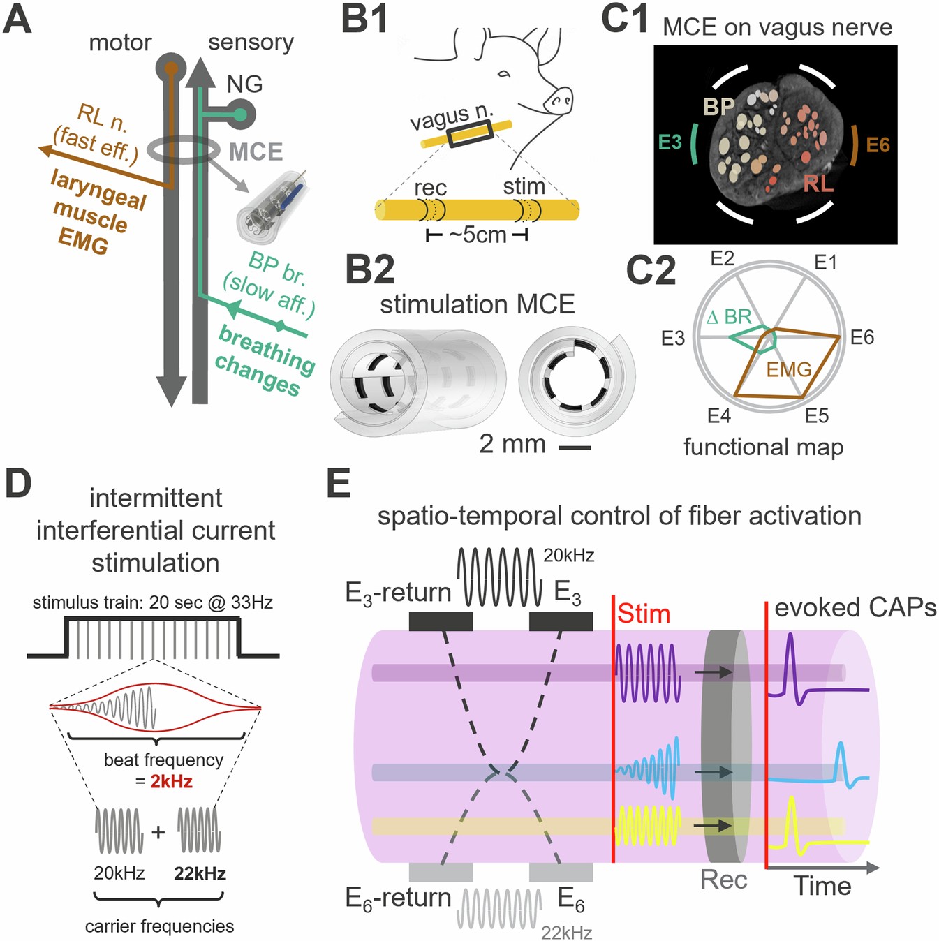
Figure 1 of Rossetti et al. 2025: vagus nerve anatomy with laryngeal and bronchopulmonary branches, a multi-contact cuff on the swine vagus nerve, a functional map of the cuff contacts, and a schematic of two kHz carriers interfering to activate selected fibers
Two kHz carriers delivered through a multi-contact cuff electrode (MCE) create a 2 kHz beat that can be steered inside the swine vagus nerve. Fig. 1 from Rossetti et al., Nature Communications 16, 4419 (2025), CC BY-NC-ND 4.0.

The problem

Conventional vagus nerve stimulation activates the whole nerve. Fibers serving the heart, lungs, larynx and other organs respond together, which limits the dose that can be given before side effects appear.

Our approach

With imec and the Zanos lab, we deliver interferential currents through multi-contact cuff electrodes, and use computational nerve models built on the ASCENT pipeline to explain what we measure in the swine vagus nerve.

Heart and breathing effects set the usable dose

Vagus nerve stimulation is usually turned up until a response appears. In rats, nerve fibers began responding at about 25 µA on average. Blood pressure and breathing changed next, at about 70 µA, followed by heart rate at about 80 µA. Implant age, electrode impedance and the type of anesthesia all shifted these thresholds.
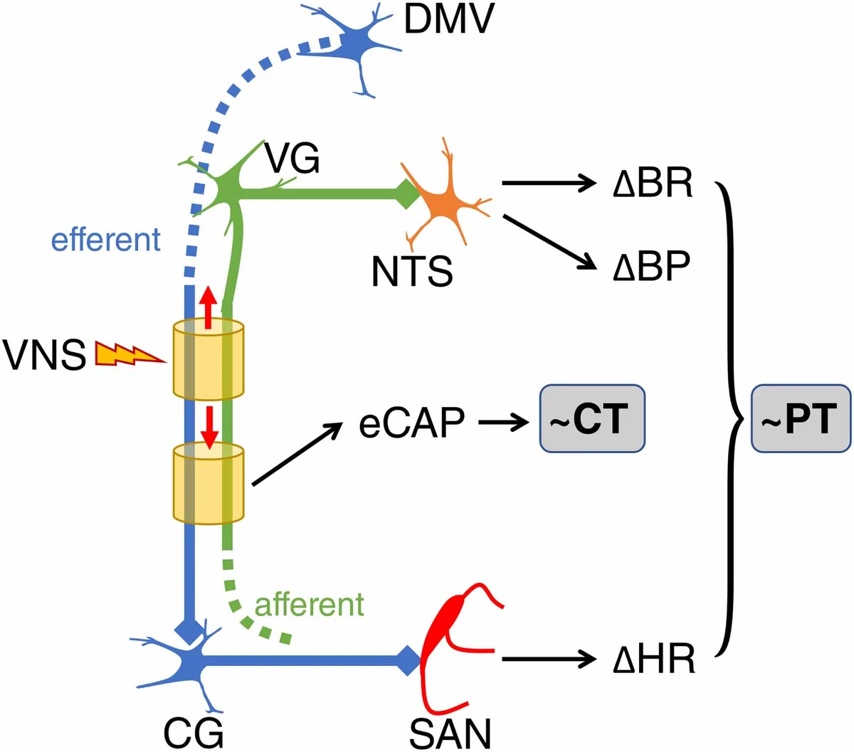
Schematic of a cuff on the vagus nerve: efferent fibers run to cardiac ganglia and the sinoatrial node (change in heart rate); afferent fibers run through the vagal ganglia to the brainstem nucleus of the solitary tract (changes in breathing and blood pressure); the evoked compound action potential defines one threshold and the physiological responses another
Efferent fibers reach the heart's pacemaker, and afferent fibers reach the brainstem, which changes breathing and blood pressure. The evoked nerve response (eCAP) defines one threshold (CT); the physiological responses define another (PT). Fig. 8 from Ahmed et al., Journal of Neural Engineering 18, 046075 (2021), CC BY 4.0.

Interfering currents can be steered across the nerve

Two high-frequency currents are delivered through different contacts of the cuff. Where they overlap inside the nerve they form a low-frequency beat, and changing the balance between the two currents moves that overlap across the nerve's cross-section.

In swine, this shifted activation between vagal fibers serving different functions, such as motor fibers to the larynx and sensory fibers from the lungs that change breathing.

kHz stimulation reaches small, unmyelinated fibers

Earlier work with the Zanos lab showed that kHz-frequency stimulation selectively activates small, unmyelinated vagus afferents, and mapped how vagal fibers are organized by organ and function inside the nerve.
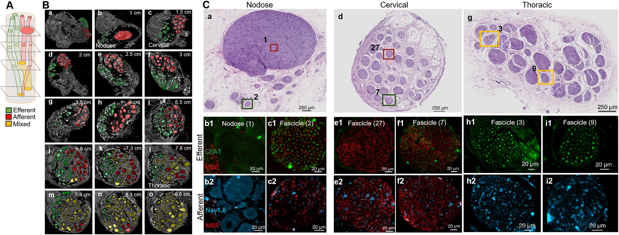
Figure with three parts: a schematic of efferent, afferent and mixed fascicles along the vagal trunk; micro-CT cross-sections at 1 to 8.6 cm from the nodose ganglion with fascicles colored green, red and yellow; and stained sections of the nodose, cervical and thoracic vagus with immunostained close-ups of individual fascicles
Tracking efferent (green), afferent (red) and mixed (yellow) fascicles along the swine vagus nerve, from the nodose ganglion to the thorax, with histology and immunostaining of individual fascicles. Full size Fig. 2 from Jayaprakash et al., Brain Stimulation 16, 484–506 (2023), CC BY-NC-ND 4.0.